Guest Seminar: Lara Wallenwein

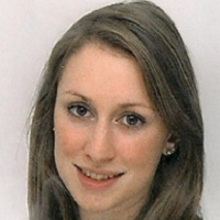
We are very happy to host Lara Wallenwein in our remote seminar series. Lara is a master's student in clinical psychology and cognitive neuroscience at the LMU Munich. The seminar will take place remotely on May 22, 2020, at 3:30 pm.
Abstract
Positron Emission Tomography (PET) using 18F-Fluorodeoxyglucose (FDG) as radiotracer is a powerful tool to assess glucose metabolism, thus allowing for a direct and quantifiable measure of neuronal activity. The bolus injection protocol is based on a steady state assumption, implying that the distribution of FDG uptake remains stable after an uptake period of approximately 30 min (Varrone et al., 2009). Controversially, a few studies found differences between subsequently acquired PET images at 60 min post-injection (p.i.) compared to 70 min p.i. (Ishii et al., 2006). To test for task-induced changes in FDG uptake, data of 49 middle-aged participants were analyzed. Imaging data were acquired within an independent research project in a hybrid PET/MR scanner at 30 to 90 min p.i. in list mode. Participants were scanned twice to analyze the effects of cognitive training intervention within the independent research project. Per scanning session two resting-state FDG-PET images representing cumulative FDG uptake from 42 min p.i. to 50min p.i (Rest1) and 50 min p.i. to 58 min p.i.(Rest2) were obtained. Additionally, one FDG-PET image representing FDG uptake whilst performing a working memory task from 60 min p.i. to 68 min p.i. (Task1) was acquired. Regional differences in FDG uptake were analyzed on a whole brain level, using a 2 x 3 ANOVA design with 2 Timepoints and 3 Brain States (i.e. Rest1, Rest2 and Task1) with global normalization. The task contrast (Task1 > Rest2) revealed mixed task- and time- induced changes in FDG uptake in frontal and parietal areas as well as in dorsal anterior cingulate cortex, and putamen. The rest contrast (Rest2 > Rest1), representing only time- induced FDG uptake variations, revealed significant differences mainly in medial frontal and temporal areas, and putamen. To isolate task-induced FDG uptake, a contrast of contrast was computed, comparing the task contrast to the rest contrast (i.e. Task1 > Rest2 >> Rest2 > Rest1). This analysis revealed significant increase in FDG uptake within clusters in frontal lobe, inferior parietal cortex, left precentral gyrus, posterior-, and dorsal anterior cingulate cortex. These are regions known to be involved in WM processing (Owen et al., 2005). Simultaneously acquired task fMRI data will be analyzed accordingly to study the overlap between fMRI and FDG-PET derived WM task activations. This study is the first to show quasi-dynamic task-induced regional changes of FDG uptake during a highly demanding cognitive task. Compared to the newly developed continuous infusion protocol for functional PET imaging (Villien et al., 2014), less FDG is required, thus increasing feasibility and decreasing radiation burden. Further studies should optimize scanning paradigms to elevate sensitivity and unfold the full potential of the procedure. From a clinical point of view, the technique may serve for diagnostic purposes, allowing the assessment of alterations in glucose metabolism during different brain states in one scanning session.
About the speaker
Lara is a master's student in clinical psychology and cognitive neuroscience at the LMU Munich, working Igor Yakushev. She is interested in multimodal imaging of normal and pathological cognition.
Details
- Date: 05/22/20
- Time: 3:30 PM CET
- Location: Remote via Zoom
- Host: Philipp Homan